Case Studies
.jpeg)
Adult
According to (Cheng et al., 2023), a horizontally impacted second molar is a rare condition that can contribute to regional malocclusion and other complications such as root resorption, caries, and periodontal pathology. This aligns with my patient’s case, as her mandibular second molar is positioned horizontally despite having undergone previous orthodontic treatment. Her case highlights the complexity of managing such impactions and supports the literature’s observation that treatment outcomes can vary depending on individual factors such as tooth inclination, age, and previous orthodontic interventions. Also according to (Sangha et al., 2021), Permanent molars erupt distal to the primary molars and can become “locked out” when there is insufficient space in the dental arch. This lack of space, known as arch length deficiency (ALD), results from a mismatch between tooth size and jaw size. ALD may occur due to disturbances in mandibular growth, abnormal tooth development, or genetic influences.
.jpeg)


History: Patient is a 21 year old female, patient does not have no significant medical history, she is in her senior year of college and has not had a cleaning in 6 months. She did have a past of orthodontic treatment for 7 years.
Vitals:
Temperature: 95.6 F
Blood Pressure:127/79 BP (taken on left arm, adult cuff)
Pulse: 66bpm normal amplitude, regular, easily felt
Respiration: 18 RPM easy respirations
EO/IO:
Face- appear symmetrically, patient has macules above left eye flat dark brown in color, 4 ear lobe piercings, skin is intact, and uniform of color, generalized little to no facial hair, no tenderness, cervix area has a flat small oval lesion macule less than 1 mm on the left side posterior, right side of the cervix area has 1 and right side has multiple black macule lesion less than 1 mm.
TMJ - No clicking or popping of the jaw as patient opens and closes, there was no asymmetrical movements observed, and patient reported no pain or tenderness.
Soft tissues of the lymph nodes: non-palpable
Submental: Non palpable
Cervical: Non-palpable
PreauriculaNon-palpableble
Post Auricular: non palpable
Submandibular left: palpable but not tender non palpable, not tender on both sides
Lips and Vermilion Border -At rest, the lips normally touch, The surface of lips is smooth and intact with a pigment color and normal texture, The vermillion border is even and not raised.
Labial/buccal mucosa- Smooth, intact, and coral pink to bluish in color, No lesions, Intact frenum on maxillary and mandibular arches. Pink with a little melanin color gingival on both arches mandibular and maxillary
gingiva: uniform pink colored pigment with little to none of melanin, fits snugly around the tooth, meets the tooth in a tapered rounded edge, texture is normal stippled, it is firm in consistent, coronal to the CEJ
Linea alba: horizontal line inside cheek
Submandibular/ Sublingual ducts: Underlying structures of lips and cheek are moist tissues, floor of mouth sublingual frenum, adequate flow of saliva, lingual frenum is very mid but seen to the eye.
Tongue - nonpalpable, lingual veins on ventral surface, bilateral scalloped edges, submental
Palate- Rugae palate is present, tori on the maxillary palatine
Oropharynx- visible
Uvula- uvula is founded at midline
OHI: Patient was instructed to floss and brush twice daily make sure she is brushing in a circular motion using the brass method and making sure she is brushing her tongue.
Dental Hygiene Diagnosis
Periodontitis stage: Stage II, Grade A
Unmet Need of Protection from Health Risks due to the presence of calculus, bleeding, and probing depths, increasing the patient’s risk for periodontal disease progression.
Goal: Patient will demonstrate improved periodontal health by flossing more daily and brush at least twice a day as indicated by reduced probing depths and bleeding.
Interventions: I plan to perform debridement and SRP as needed.
I will schedule patient to come back to check the progress for regular 3–4 month periodontal maintenance intervals.
Monitor periodontal status with periodontal charting and radiographs.
Unmet Need of Freedom from Fear and Stress due to possible emotional stress from prolonged orthodontic treatment (7 years) and current flared anterior teeth (#8–9, #24–25).
Goal: Patient will report reduced anxiety and increased comfort during dental visits.
Interventions: Use patient-centered communication and reassure the patient about procedures.
Encourage the patient to express concerns and esthetic goals.
I will offer an referral from Dr. Forester to an orthodontist oral surgeon, as appropriate.
Unmet Need of Freedom from Pain due to horizontal impaction of both mandibular second molars, potentially causing discomfort.
Goal: Patient will remain free of pain and understand the need for oral surgery consultation.
Interventions: Dr. Forester will conduct an referral for the patient to oral surgeon for evaluation of impacted teeth.
Monitor area for symptoms of pain.
Educate patient on signs of infection and when to seek care.
Unmet Need of Wholesome Facial Image due to flared anterior teeth resulting from or persisting after orthodontic treatment, possibly impacting self image.
Goal: Patient will express satisfaction with facial appearance or pursue consultation for esthetic correction.
Interventions: Provide education on available esthetic options (orthodontic retreatment, cosmetic restorations).
Offer referral to orthodontist or cosmetic dentist.
Provide emotional support regarding facial aesthetics.
Unmet Need of Skin and Mucous Membrane Integrity of the Head & Neck due to oral hygiene of not flossing regularly evidenced by generalized bleeding on probing (BOP), probing depths of 4mm, and slight calculus build up.
Goal: Patient will reduce gingival inflammation and bleeding within 4–6 weeks as demonstrated by a decrease in BOP and improved gingival health.
Interventions: Perform full mouth debridement and SRP as needed.
Educate patient on proper brushing technique (Modified Bass) and daily interdental cleaning such as using a waterpik from Amazon, Walmart, Target, or a daily interdental flosser an suggestion is the "Gum proxabrush go betweens" at Walmart
Recommend antimicrobial rinse (e.g., chlorhexidine) to control bacteria.
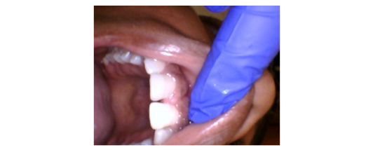
Unmet Need of Biologically Sound and Functional Dentition due to suboptimal plaque control and possible dietary habits, evidenced by a suspicious carious lesion on tooth #12.
Goal: Patient will prevent progression of carious lesion on #12 through improved self-care and dietary habits, and seek appropriate restorative treatment.
Interventions: Refer patient to dentist for evaluation and possible restoration of #12.
I will provide dietary counseling to reduce frequency of sugar intake.
Reinforce use of fluoride toothpaste and recommend "Sensodyne, Repair and Protect"
Unmet Need of Conceptualization and Problem Solving due to a more understanding of periodontal status and options for care, as evidenced by calculus buildup and probing depths.
Goal: Patient will verbalize understanding of current oral condition and treatment plan.
Interventions: Use visual aids and radiographs to explain findings. Offer Q&A and show tell do method during during appointments to clarify misconceptions. Provide educational materials tailored to the patient’s learning preferences.
Unmet Need of Responsibility for Oral Health due to insufficient daily plaque removal and resulting gingival inflammation and calculus accumulation.
Goal: Patient will demonstrate improved oral hygiene practices and plaque control.
Interventions: Provide tailored oral hygiene instruction, including brushing and interdental aids.
Set short-term hygiene goals (reduced plaque scores by next visit).
Reinforce motivation and accountability through follow-up and encouragement.
Adolescent
According to (National Institute of Dental and Craniofacial Research(US), 2021) Heritably and genetics play an important role in the wide spectrum of malocclusions, but environment and oral habits also are critical factors in the dental and facial variations observed in children or adolescents. According to the oral health surveillance report in 2011–2016 from CDC, more than half (57%) of adolescents aged 12–19 years experienced dental caries. One in six adolescents aged 12–19 years had untreated tooth decay (Zhang et al., 2023b)
History: Patient is a 12 year old female, healthy patient no systemic diseases. Patient has orthodontic treatment and does not have no chief complaints. Patient last cleaning was 6 months ago. She does have a dental home which is in Cincinnati, OH where she is from.
Vitals:
Temperature: 95.5
Pulse: 60 BPM
Respiration: 24 RPM
Blood Pressure: 110/68 mmHg (taken on the left arm using the pedo cuffs)
Occlusion:
Molar right Relationship: Class I relationship Class I canine
Left Molar Relationship: Class I relationship Class I canine
Anterior Overjet: slight 2mm
Anterior overbite: slight
Anterior Edge to Edge: N/A
Anterior Open Bite: N/A
Anterior Cross Bite: N/A
Posterior End to End: N/A
Posterior Crossbite: N/A
Malposition of teeth: N/A
Other findings: N/A
8,9- facially slight protruded
24,25- flat protruded
EO/IO:
Facial appearance: face appear asymmetrically, hyperpigmentation on forehead, acne visual on forehead also hair is prominent on forehead and around side burns. Skin was smooth, intact.
Temporomandibular Joint (TMJ): Palpation of bilateral TMJs revealed no tenderness, joint noise (clicking, popping) or deviation.No limitations or deflection during opening, closing.
Lymph Nodes:
Occipital: Not palpable and nontender, no swelling or firmness noted.
Postauricular: Not palpable, nontender bilaterally, no indication of ear infection. flat macule by ear lobe, ear lobe are pierced on both sides
Preauricular: Symmetrical, nonpalpable, and nontender, no signs of localized infection or inflammation
Submandibular: nontender and nonpalpable
Submental: Small, mobile, and nontender. No signs of inflammation associated with mandibular anterior, lower lip, or chin area.
Cervical Chain (Anterior and Posterior): Both anterior and posterior chains were nonpalpable and nontender. No signs of respiratory infection.
Supraclavicular: Flat, nontender, and nonpalpable bilaterally.
Salivary Glands: Submandibular Glands: non tender and nonpalpable submandibular within normal limits
Sublingual Glands: Noted intraorally under the tongue. Adequate saliva observed with no obstruction or pain.
Thyroid Gland: No nodules or enlargement observed, No tenderness reported on swallowing
Lips:
Lips were well hydrated, melanin with slight pink, and smooth. Vermillion borders were intact and well defined. No cracking was present. No lesions or evidence of lip biting from ortho brackets.
Labial Mucosa:
Appeared pink, moist, and intact. No trauma or ulcerations use noted, Maxillary and mandibular labial frena were normally attached with no abnormal pull
Submandibular/ Sublingual ducts: Underlying structures of lips and cheek are moist tissues, floor of mouth sublingual frenum, adequate flow of saliva, lingual frenum is very mid but seen to the eye.
Buccal Mucosa:
Bilateral buccal mucosa were smooth and pink, A bilateral Linea alba was present, likely due to habitual cheek biting or buccal contact from orthodontic appliances.
Gingiva:Attached gingiva was coral pink with localized physiologic melanin pigmentation in the anterior, Mild generalized inflammation noted around bracketed areas, primarily in the maxillary and mandibular anterior regions.
Tongue: Dorsal Surface: Pink with a thin, uniform white plaque coating likely from biofilm accumulation, not candidiasis. trauma present under tongue is present on the right side.
Lingual Frenum: Intact
Floor of the Mouth:
Moist, pink, and smooth, Sublingual folds and frenum were intact and within normal limits, ducts visible bilaterally with clear salivary flow on palpation, Mandibular tori were present bilaterally, more prominent on the anterior. Firm and covered with healthy mucosa.
Hard Palate: Pale pink and firm to palpation. Rugae were symmetrical and well defined, No lesions, ulcerations and palatal tori is present.
Soft Palate and Uvula: Soft palate appeared pink, smooth, and healthy, Uvula was midline, of normal size, and moved symmetrically on phonation.
Oropharynx & Tonsils: Posterior pharyngeal wall was pink, moist, Tonsils were present and not enlarged


Perio Case
The period of young adulthood (ages 15–49) represents a crucial yet often under researched stage marked by the development of metabolic risk factors such as diabetes and hypertension. These conditions can increase susceptibility to periodontal diseases. Other contributing factors include smoking, obesity, poor diet, and stress all of which are strongly linked to the lifestyle and behavioral patterns commonly seen in young adults (Wang et al., 2025). Periodontitis is a bacterial gum infection that damages the soft tissue and supporting structures of the teeth. Arestin, an antibiotic medication, is used as an adjunctive treatment to help manage periodontitis by targeting and eliminating the bacteria responsible for the infection. By reducing bacterial levels, Arestin helps decrease inflammation and swelling, allowing the gum tissue to heal and reattach more effectively to the tooth surface (Arestin [Minocycline]: Uses, Side Effects, Interactions, Pictures, Warnings & Dosing - WebMD, n.d.).


Statement of Health: 21 year old female patient, ASA Classification II, mild systemic disease (Rheumatoid Arthritis) only takes medication when needed, patient states she takes ibuprofen when it flares up and it rarely flares up. Hospitalized more than 4 years ago for appendix, no significant findings in dental health, last dental visit was more than 3 years ago.
Vitals:
Temperature: 97.2
Blood Pressure: 114/72 mmHg (taken on the left arm using the adult cuff)
Pulse: 60 BPM
Respiration: 24 RPM
Extraoral: WNL
Intraoral: WNL
Periodontal charting: BOP, localized 5mm
Radiographic findings: horizontal bone loss
Plaque index / calculus presence: PCR: 40%
Periodontal status (AAP classification): Stage II Grade A
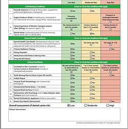
Caries risk assessment: Moderate
Occlusion:Right molar Occlusion: Class I occlusionRight Canine Occlusion: Class I occlusionLeft molar occlusion: Class I malocclusionLeft Canine Occlusion: Class I occlusionAnterior Overbite: Severe (deep)Anterior Overjet: 3mmAnterior Edge to Edge: N/AAnterior Open Bite: N/AAnterior Crossbite: N/APosterior End to End: N/APosterior Crossbite: N/A
EO/IO:
Head and Neck:
Facial apperance is asymmetrical aligned, patient has acne and hyperpigmentation located around the cheeks of the face, patient does have a tattoo located on the post auricular lymph node on the right side. Nasal piercing on left side and septum piercing.
Lymph Nodes:
Occipital: Nonpalpable non tender and patient does have a scratch in the middle of the neck, but no swelling.
Postauricular nodes: patient does have a red ink heart shaped tattoo but patient states nontender and nonpalpable, ear lobe are pierced on both sides
Preauricular nodes: Nonpalpable nontender, no signs of localized infection or inflammation
Submental nodes: mobile on the right side of the mandibular, nontender, no signs of inflammation associated with the mandibular anterior.
Submandibular nodes: patient stated uncomfortable but not in pain due to the third molar being uncomfortable also because radiology is showing the third molar growing in horizontally.
Cervical Nodes (Anterior and Posterior): Both anterior and posterior chains were nonpalpable and nontender. No signs of respiratory infection.
Supraclavicar nodes: Flat, nontender, and nonpalpable bilaterally
TMJ: No clicking or popping at jaw, no limitations or deflection during opening and closing
Submandibular glands: non tender and nonpalpable submandibular within normal limits
Sublingual Glands: Noted intraorally under the tongue. Adequate saliva observed with no obstruction or pain.
Thyroid Gland: No nodules or enlargement observed, No tenderness reported on swallowing
Lips: Lips were well hydrated, melanin with slight pink, and smooth. Vermillion borders were intact and well defined. No cracking was present.
Labial Mucosa: Appeared pink, moist, and intact.
Submandibular/ Sublingual ducts: structures of lips and cheek are moist tissues, adequate flow of saliva
Buccal Mucosa: Bilateral buccal mucosa were smooth and pink, A bilateral Linea alba was present on right and left side
Gingiva: Attached gingiva was coral pink with melanin pigmentation in the anterior
Tongue: Pink with a thin, uniform white plaque coating likely from biofilm accumulation, not candidiasis.
Lingual Frenum: Intact
Floor of the Mouth: Moist, pink, and smooth, Sublingual folds and frenum were intact and within normal limits but mild to see to the eye, ducts visible bilaterally with clear salivary flow on palpation, Mandibular tori were present bilaterally, more prominent on the anterior. Firm and covered with healthy mucosa.
Hard Palate: Pale pink and firm to palpation. Rugae were symmetrical and well defined, No lesions, ulcerations and small palatal tori is present midline.
Soft Palate and Uvula: Soft palate appeared pink, smooth, and healthy, Uvula was midline, of normal size
Oropharynx/ Tonsils: Posterior pharyngeal wall was pink, moist, Tonsils were enlarged.
Older Adult
Statement of Health: 68-year-old female patient, ASA Classification III, moderate systemic disease (controlled Type 2 Diabetes and Hypertension). Patient reports taking Metformin 500 mg twice daily and Lisinopril 10 mg daily as prescribed. Last A1C reported at 7.1%. Hospitalized 6 years ago for knee replacement surgery with no complications. Patient states last dental visit was more than 8 years ago. Reports occasional bleeding gums, tooth mobility, and sensitivity while chewing.
Vitals:
Temperature: 98.1°F
Blood Pressure: 138/84 mmHg (taken on the left arm using the adult cuff)
Pulse: 76 BPM
Respiration: 20 RPM
Extraoral: WNL
Intraoral: Generalized inflammation noted
Periodontal charting: Generalized BOP, multiple 6–9 mm periodontal pockets, recession present, furcation involvement on molars, class II mobility on mandibular anterior teeth
Radiographic findings: Generalized severe horizontal and vertical bone loss extending to middle/apical third of roots, calculus deposits visible radiographically, furcation involvement present
Plaque index / calculus presence: PCR: 82%, heavy generalized supragingival and subgingival calculus
Periodontal status (AAP classification): Stage IV Grade C generalized periodontitis
Caries risk assessment: High
Occlusion:
Right molar Occlusion: Class II occlusion
Right Canine Occlusion: Class II occlusion
Left molar occlusion: Class II occlusion
Left Canine Occlusion: Class II occlusion
Anterior Overbite: Moderate
Anterior Overjet: 5 mm
Anterior Edge to Edge: N/A
Anterior Open Bite: N/A
Anterior Crossbite: N/A
Posterior End to End: N/A
Posterior Crossbite: N/A
EO/IO:
Head and Neck:
Facial appearance is symmetrical and aligned. Mild wrinkling consistent with age. No visible lesions. Patient wears prescription glasses. No facial swelling noted.
Lymph Nodes:
Occipital: Nonpalpable, nontender, no abnormalities detected.
Postauricular nodes: Nonpalpable, nontender, no localized infection present.
Preauricular nodes: Nonpalpable, nontender, no swelling observed.
Submental nodes: Palpable but mobile and nontender, no associated inflammation.
Submandibular nodes: Slightly enlarged bilaterally, mobile and mildly tender due to active periodontal inflammation.
Cervical Nodes (Anterior and Posterior): Nonpalpable and nontender. No signs of infection.
Supraclavicular nodes: Flat, nontender, and nonpalpable bilaterally.
TMJ: Mild crepitus on opening, no pain reported, no limitations in movement.
Submandibular glands: Nontender and within normal limits.
Sublingual Glands: Visible intraorally, reduced salivary flow noted.
Thyroid Gland: No enlargement or nodules observed.
Lips: Dry with mild cracking at commissures, otherwise intact.
Labial Mucosa: Pink, moist, no lesions present.
Submandibular/Sublingual ducts: Reduced but adequate salivary flow.
Buccal Mucosa: Pink with slight generalized dryness, bilateral linea alba present.
Gingiva: Generalized erythematous, edematous gingiva with rolled margins, recession present throughout posterior and anterior sextants. Heavy plaque and calculus accumulation observed along cervical margins.
Tongue: Pink with moderate fissuring and thin white coating consistent with biofilm accumulation.
Lingual Frenum: Intact.
Floor of the Mouth: Pink, smooth, and moist. Ducts visible bilaterally. No lesions observed.
Hard Palate: Pale pink, firm, no lesions present.
Soft Palate and Uvula: Pink, smooth, healthy appearance, uvula midline.
Oropharynx/Tonsils: Posterior pharyngeal wall pink and moist. Tonsils within normal limits for age.
Pediatric
Statement of Health: 8-year-old female patient, ASA Classification I, no known systemic disease. Patient is not currently taking any medications. Parent reports no history of hospitalization or surgical procedures. Last dental visit was over 2 years ago. Parent reports occasional gum bleeding during brushing and frequent snacking on sugary foods and juices.
Vitals:
Temperature: 98.6°F
Blood Pressure: 98/60 mmHg (taken on the left arm using the pediatric cuff)
Pulse: 92 BPM
Respiration: 20 RPM
Extraoral: WNL
Intraoral: Mild generalized inflammation noted
Periodontal charting: Generalized BOP, localized 5–6 mm pockets on erupting permanent first molars, pseudopocketing present due to gingival enlargement
Radiographic findings: Localized horizontal bone loss around erupting permanent first molars, radiographic calculus deposits noted on mandibular anterior teeth
Plaque index / calculus presence: PCR: 72%, moderate to heavy supragingival calculus on mandibular anterior teeth, generalized plaque accumulation
Periodontal status (AAP classification): Stage III Grade C localized periodontitis
Caries risk assessment: High
Occlusion:
Right molar Occlusion: Flush terminal plane
Right Canine Occlusion: Class I tendency
Left molar occlusion: Flush terminal plane
Left Canine Occlusion: Class I tendency
Anterior Overbite: Moderate
Anterior Overjet: 3 mm
Anterior Edge to Edge: N/A
Anterior Open Bite: N/A
Anterior Crossbite: N/A
Posterior End to End: N/A
Posterior Crossbite: N/A
EO/IO:
Head and Neck:
Facial appearance is symmetrical and aligned. No swelling or visible lesions. Mild freckles across nasal bridge and cheeks.
Lymph Nodes:
Occipital: Nonpalpable, nontender.
Postauricular nodes: Nonpalpable, nontender.
Preauricular nodes: Nonpalpable, nontender.
Submental nodes: Palpable, mobile, and nontender.
Submandibular nodes: Slightly enlarged, mobile, and mildly tender due to localized oral inflammation.
Cervical Nodes (Anterior and Posterior): Palpable anterior cervical nodes, mobile and nontender, likely related to mild chronic inflammation.
Supraclavicular nodes: Nonpalpable and nontender.
TMJ: No clicking, popping, or pain. Full range of motion.
Submandibular glands: Within normal limits.
Sublingual Glands: Adequate salivary flow observed.
Thyroid Gland: No enlargement or abnormalities noted.
Lips: Pink, hydrated, and intact.
Labial Mucosa: Pink and moist, no lesions.
Submandibular/Sublingual ducts: Clear salivary flow bilaterally.
Buccal Mucosa: Smooth and pink, faint bilateral linea alba present.
Gingiva: Generalized erythema with edematous margins, localized calculus deposits on mandibular anterior teeth, plaque biofilm visible along gingival margins.
Tongue: Pink with light coating on dorsum.
Lingual Frenum: Intact.
Floor of the Mouth: Pink, moist, and smooth.
Hard Palate: Pale pink and firm, no abnormalities noted.
Soft Palate and Uvula: Healthy appearance, uvula midline.
Oropharynx/Tonsils: Posterior pharyngeal wall pink and moist, tonsils slightly enlarged bilaterally.